
In addition to allowing precise, close-up work, the microscope directs a beam of light directly on the teeth, minimizing glare for you.
Many dentists use air-driven “handpieces” (the dental term for “drills”). While acceptable for many procedures, these “whiney-sounding” air-powered handpieces all have a degree of non-concentricity; they do not rotate perfectly smoothly.
For the most precise aspects of restorative procedures, we use electric handpieces. This results in extraordinarily precise interfaces between your tooth and your new restoration (dental crown, tooth veneer, or tooth filling). This will bring more comfort to you as well. With a more precise tool, there is less vibration and less noise.
Digital Imaging
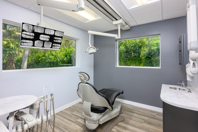
Drs. Warner or Scoglio choose carefully which and when radiographs are taken. There are many guidelines that we follow. Radiographs allow us to see everything we cannot see with our own eyes. Radiographs enable us to detect cavities between your teeth, determine the bone level, and analyze the health of your bone.We can also examine the roots and nerves of teeth, diagnose lesions such as cysts or tumors, as well as assess damage when trauma occurs.
Dental radiographs are invaluable aids in diagnosing, treating, and maintaining dental health. Exposure time for dental radiographs is extremely minimal. Drs. Warner and Scoglio utilize Digital Imaging Technologies within the office. With digital imaging, exposure time is about 50 percent less when compared to traditional radiographs. Digital imaging can also help us retrieve valuable diagnostic information. We may be able to see cavities better.
Digital imaging allows us to store patient images, and enables us to quickly and easily transfer them to specialists or insurance companies.
Digital X-Rays:
Digital X-rays offer more precision since we view the image on a computer monitor, instead of holding up a 35mm film up to the light. Digital X-rays results in 1/6th the radiation exposure to you.
iTero® Intraoral 3D Scanner
The iTero Intraoral 3D Scanner is an advanced digital imaging system that allows our team to create highly accurate, 3D digital models of your teeth and gums—without the mess or discomfort of traditional impressions. Using a small handheld wand, we gently scan your mouth to capture thousands of detailed images in just minutes. These images are then assembled into a precise 3D model that we can view instantly on screen.
This technology eliminates the need for trays filled with impression material, providing a faster, cleaner, and more comfortable experience for our patients. The iTero system is safe, radiation-free, and ideal for patients of all ages.
Common Uses of the iTero Scanner
- Invisalign® and Clear Aligner Treatment: We use iTero scans to plan and monitor orthodontic treatment. The digital model helps us design aligners that fit perfectly and even simulate your “after” smile before treatment begins.
- Crowns, Bridges, and Veneers: iTero scans provide the precision needed to create restorations that fit seamlessly and look natural—often eliminating the need for physical impressions or multiple appointments.
- Implant Planning: The detailed 3D data helps us plan implant placement with exceptional accuracy and visualize how restorations will align with your natural bite.
- Diagnostic Records: Because digital scans are stored electronically, we can track changes in your teeth and gums over time with remarkable precision.
Benefits to Patients
- Fast, comfortable, and impression-free experience
- Instant visualization of your treatment plan
- More precise and predictable results
- Environmentally friendly—no disposable impression materials
With iTero digital scanning, your dental visits are more efficient and comfortable, and your restorations and aligners are designed with greater accuracy than ever before.
Intraoral Camera
Many patients, especially younger patients, are very familiar with the latest technology and are comfortable with the high-tech practice. Computers and TV screens are their primary method of information processing.
Drs. Warner and Scoglio utilize Intraoral Camera technology that helps enhance your understanding of your diagnosis. An Intraoral Camera is a very small camera – in some cases, just a few millimeters long. An Intraoral Camera allows our practice to view clear, precise images of your mouth, teeth, and gums, in order for us to accurately make a diagnosis. With clear, defined, enlarged images, you see details that may be missed by standard mirror examinations. This can mean faster diagnosis with less chair-time for you!
Intraoral cameras also enable our practice to save your images in our office computer to provide a permanent record of treatments. These images can be printed for you, other specialists, your lab, or insurance companies.